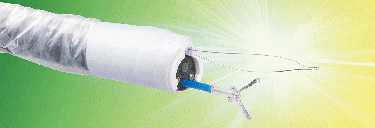
In lesions > 2 cm, EMR+ outdoes its advantages: comparative trial demonstrates en-bloc resection rate of 86.36 % in 3 cm lesions with EMR+ vs. 18.18 % with conventional EMR
For larger sessile or laterally spreading gastrointestinal polyps ≥ 2 cm, en-bloc resection with the conventional EMR technique is technically very difficult. EMR with a piecemeal technique can be performed but this is associated with higher recurrence rates. The classical EMR technique can be improved with a new technique using an external additional working channel (AWC) that is mounted on a standard endoscope similar to the setup used with the FTRD® System. The technique is called EMR+.
Knoop RF et al., Department of Gastroenterology and Gastrointestinal Oncology, University Medical Center, Goettingen, Germany, evaluated the EMR+ technique prospectively in flat lesions comparing it with a conventional EMR. The study was conducted in an ex vivo animal model with porcine stomachs placed into the EASIE-R simulator. Prior to intervention, standardized lesions were set by coagulation dots, measuring 1 cm (n = 12 per group), 2 cm (n = 22 per group), 3 cm (n = 22 per group), or 4 cm (n = 20 per group).
Overall, 152 procedures were performed. In lesions of 1 cm, both EMR and EMR+ were very reliable with en-bloc resection rates (R0) of 100 %. In 2 cm lesions, EMR+ en-bloc resection rate was significantly higher (p = 0.02), conventional EMR already dropped to 54.55 % en-bloc resection rate, while EMR+ still achieved an en-bloc resection rate of 95.44 %. Conventional EMR did not provide sufficient resection rates for lesions of 3 cm or even 4 cm (18.18 % and 0 %). EMR+ still achieved very satisfying results in 3 cm lesions (86.36 %, p < 0.01) but also relevantly decreased at 4 cm (60.00 %, p < 0.01). From 3 cm on, EMR+ was significantly faster than conventional EMR. In 3 cm lesions, procedural time was 5 min with EMR+ vs. 12.5 min with conventional EMR, p < 0.01. In 4 cm lesions, procedural time was 5.5 min with EMR+ vs. 15 min with conventional EMR, p < 0.01.
In lesions of all sizes, the resection area was significantly larger in the EMR+ groups. In 1 cm lesions, the median resection area was 4.44 cm2 for EMR+ vs. 3.14 cm2 for conventional EMR, p = 0.012. In 2 cm lesions, it was 5.94 cm2 for EMR+ vs. 3.30 cm2 for conventional EMR, p < 0.01. At 3 cm it was 9.62 cm2 for EMR+ vs. 1.50 cm2 for conventional EMR, p < 0.01. In 4 cm lesions, EMR+ median resection area was 13.37 cm2 vs. 4.02 cm2 for conventional EMR, p = 0.03. A perforation rate of 15 % (3/20) was observed in the 4 cm-group treated with EMR+.
The authors summarized that EMR+ enables a grasp-and-snare technique and consequently facilitates en-bloc resection of larger lesions compared to conventional EMR. In lesions > 2 cm, EMR+ outdoes its advantages, especially concerning en-bloc resection rate. At 3 cm, EMR+ reaches its best discriminatory power. At 4 cm, also EMR+ comes to its inherent limits and the risk of perforations rises. Then, ESD or surgery should be considered. The authors concluded that EMR+ could help to close a therapeutic gap in interventional endoscopy with manageable technical complexity, time and costs.
Endoscopic mucosal resection with an additional working channel (EMR+) in a porcine ex vivo model: a novel technique to improve en-bloc resection rate of snare polypectomy
Knoop RF, Wedi E, Petzold G, Bremer SCB, Amanzada A, Ellenrieder V, Neesse A, Kunsch S.
Endoscopy International Open 2020; 08: E99-E104. https://doi.org/10.1055/a-0996-8050


 Deutsch
  Français