
 Deutsch
  Français
  Español
  Português
 Application aid for mobilisation of the anastomosis sides


+49 (0) 7071 96528 160
service@ovesco.com
+49 (0) 7071 96528 160
service@ovesco.com
Mobilisation aid for bringing together the two opposing anastomosis sides in the BARS® application cap.
The BARS® Anchors are supplied in black and silver so they can be easily differentiated.
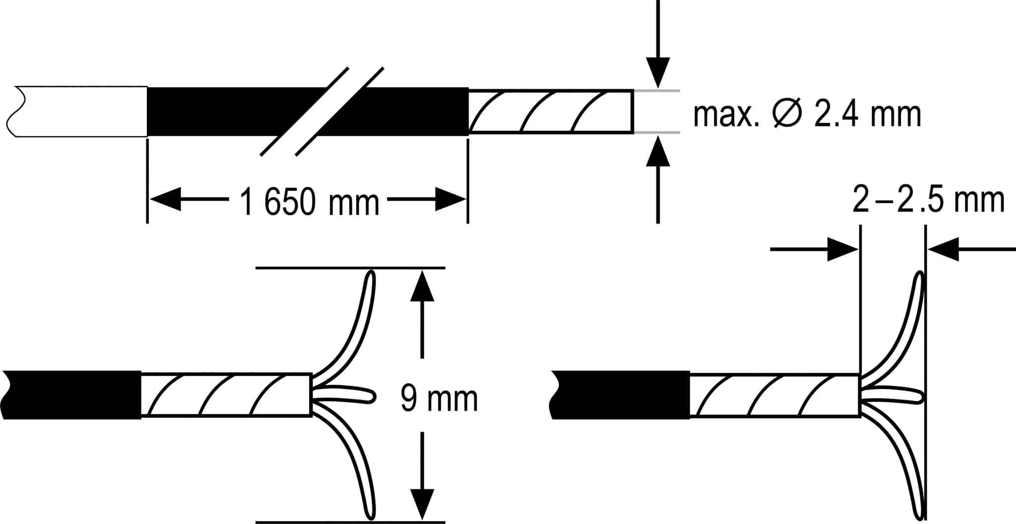
| Colour | Black and silver |
| Working length | 1650 mm |
| Max. outer diameter | 2.4 mm |
| Needle width | 9 mm |
| Stitch depth | 2-2.5 mm |
| Reference number | BARS® Set: 100.60 (incl. BARS® Anchors) |
EMR2

Mucosal incision2
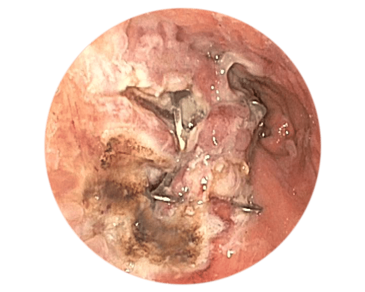
Preparing the target tissue
Preparing the target tissue using EMR or mucosal
incision offers significant benefits.
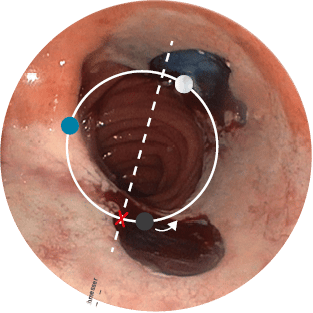
Positioning
Correct positioning of the BARS® Anchors is crucial to the success of the treatment.
2Source: Prof. A. Schmidt, Universitätsklinikum Freiburg, Germany
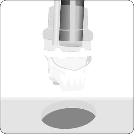
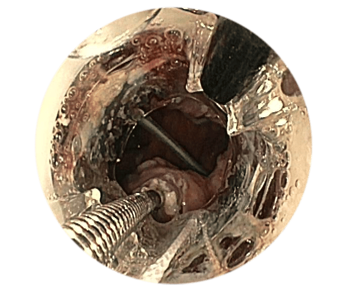
Target the application site.
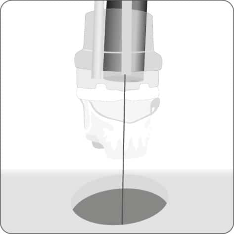
Insert the guide wire into the outer BARS® working channel and place it in the anastomosis.
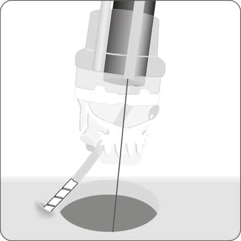
Insert the anchors into the endoscope working channel and the inner
BARS® working channel.

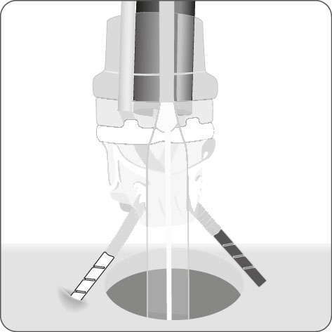
Crossed placement of the anchors in the prepared target tissue.
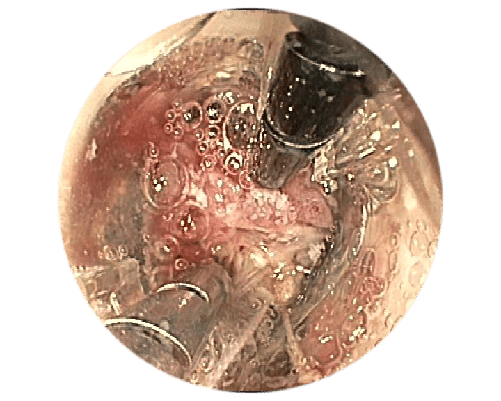
Place the calibration balloon in the anastomosis. Gradually pull the tissue into the BARS® application cap using alternating pulling motions.
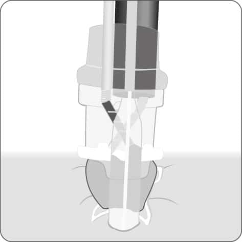
Make sure that the tissue is positioned symmetrically in the cap. Position the BARS® clip by turning the hand wheel.

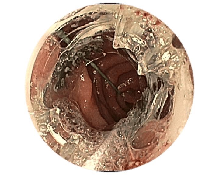
Withdraw the instruments and inspect the clip application site.

Target the application site.
Insert the guide wire into the outer BARS® working channel and place it in the anastomosis.
Insert the anchors into the endoscope working channel and the inner
BARS® working channel.

Crossed placement of the anchors in the prepared target tissue.
Place the calibration balloon in the anastomosis. Gradually pull the tissue into the BARS® application cap using alternating pulling motions.
Make sure that the tissue is positioned symmetrically in the cap. Position the BARS® clip by turning the hand wheel.
Withdraw the instruments and inspect the clip application site.
1Source: Dr. med. M. Kandler, Städtisches Klinikum Dresden, Germany
