
 Deutsch
  Français
  Español
  Português
 Innovative clipping system intended for endoscopic treatment of gastrointestinal hemorrhage and closure of acute and chronic wall lesions.


+49 (0) 7071 96528 160
service@ovesco.com
+49 (0) 7071 96528 160
service@ovesco.com
The OTSC® System Set is an instrument for flexible endoscopy. It can be used for compression of tissue in the gastrointestinal (GI) tract, for hemostasis or for treating gastrointestinal organ wall lesions, and for marking of lesions. The clip OTSC® gc is specially designed for the treatment of perforations and lesions of the stomach.
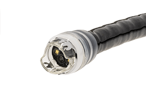
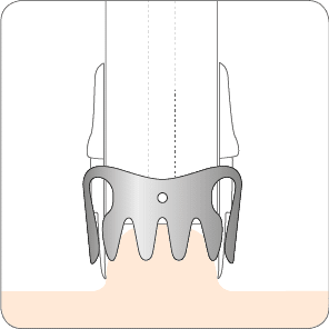
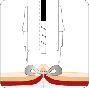
The OTSC® System Set consists of an applicator cap with a mounted OTSC® clip, thread, thread retriever and a hand wheel for clip release.
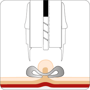
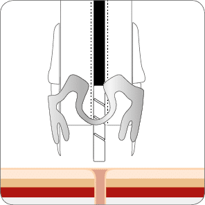
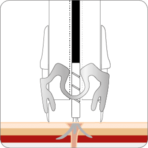
The OTSC® clip is delivered by means of an applicator cap mounted to the tip of gastroscopes or colonoscopes. The clip is released by tightening the thread with the hand wheel.
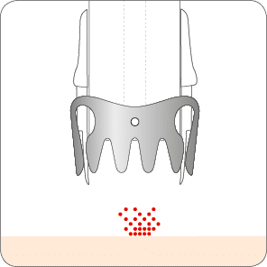
The OTSC® clip for flexible endoscopy is a superelastic Nitinol device for compression and approximation of tissue in the digestive tract.
Based on its unique design the clip closes itself and firmly anchors the tissue to be compressed for hemorrhage or closure of a GI organ wall lesion. Due to its smart material properties, OTSC® clip delivers constant force at the implantation site securing the therapeutic effect. The OTSC® clip is made of a biocompatible and MR conditional material and can remain in the body as a longterm implant.
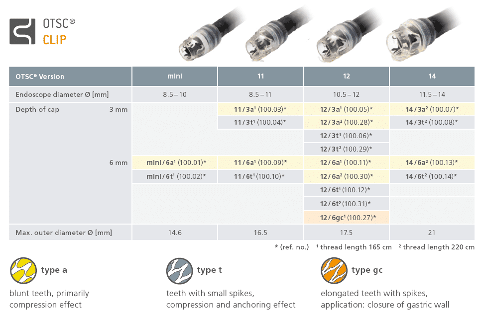
The OTSC® System is very flexible and will thus allow for multiple indications and most of the conventional equipment in endoscopy. There are various clips and caps classified as follows:
Features and sizes of the applicator caps:
Features of the clips:
The article numbers can be found in the table below or in our reference list.
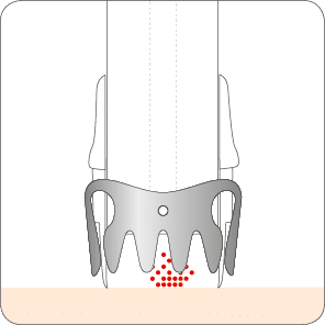
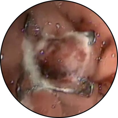
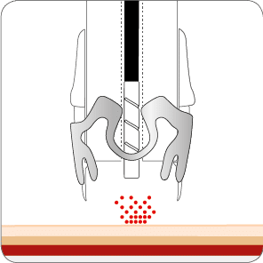
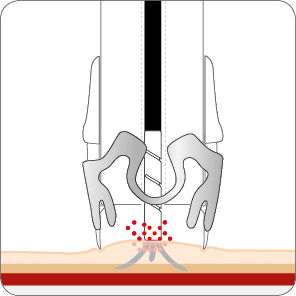
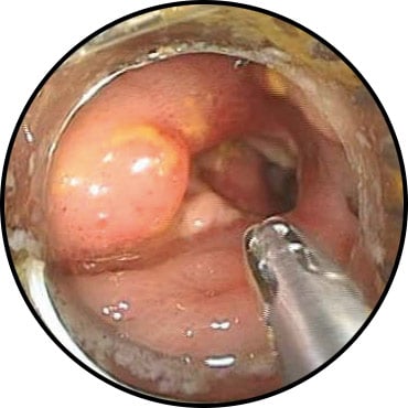
In most GI bleeding situations, tissue can be mobilized and securely pulled inside the application cap by simply applying endoscopic suction. Once the target tissue is captured inside the cap, hemostasis is achieved by turning the handwheel to release the OTSC® clip around the captured tissue.

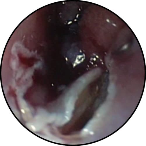
Hemostasis of arterial bleeding
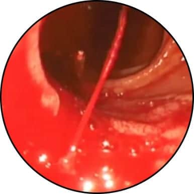
* Source: Prof. Dr. Chiu, Prince of Wales Hospital, Hong Kong SAR, China
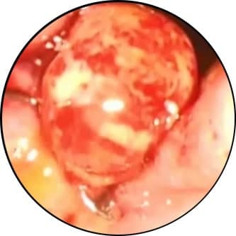
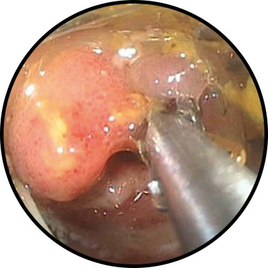
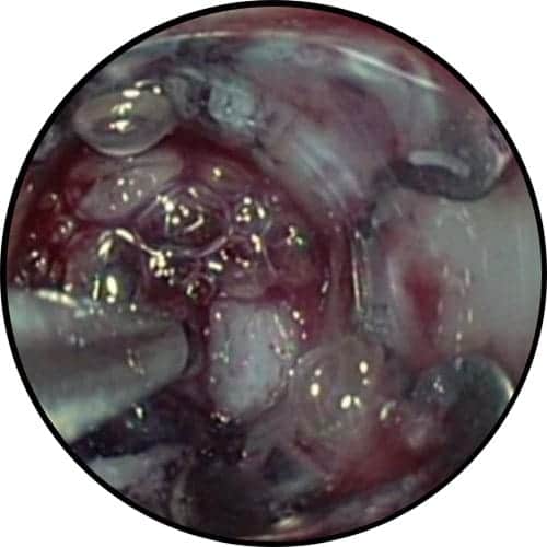
Bleeding peptic ulcer in the gastric antrum (anticoagulated patient)
* Source: Dr. Thomas Kratt, Interventional Endoscopy, Klinik für Allgemeine, Viszeral- und Transplantationschirurgie, University Hospital Tuebingen, Germany
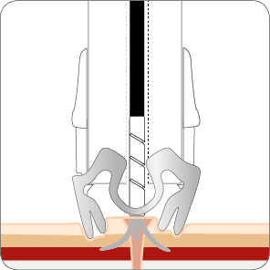
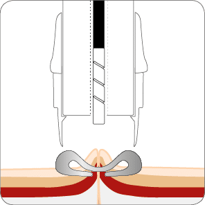
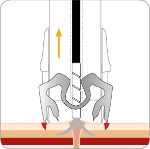
In cases of fibrotic or hard tissue (e.g. callous ulcers) or tangential application, the OTSC® Anchor can be valuable in precisely aligning target tissue with the cap opening and keeping it fixed during clip release. It may not always be possible to manipulate fibrotic tissue fully inside the cap. However, it is sufficient to pull the tissue firmly to the rim of the cap with the OTSC® Anchor, then apply the clip. The clip “jumps” slightly forward upon release and grasps the tissue in front of the cap.

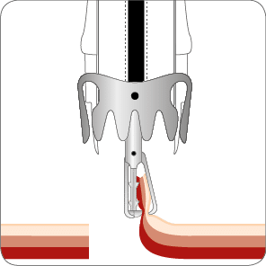
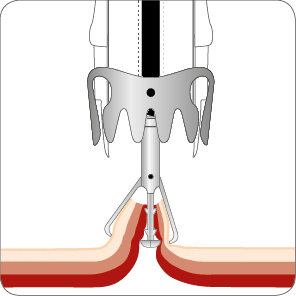
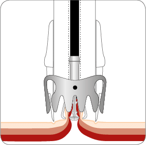
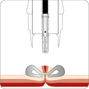
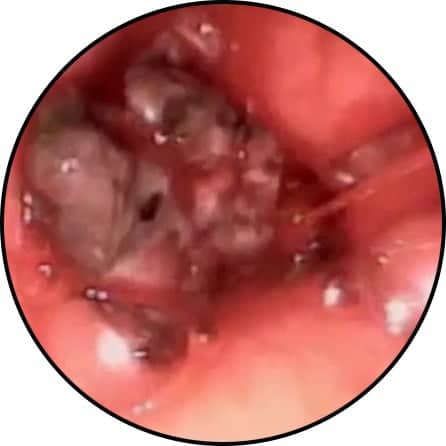
Perforation closure in the colon with OTSC® Twin Grasper®


* Source: Dr. Thomas Kratt, Interventional Endoscopy, Klinik für Allgemeine, Viszeral- und Transplantationschirurgie, University Hospital Tuebingen, Germany
Closure of persistent PEG-fistula using the OTSC® Anchor


* Source: Dr. Thomas Kratt, Interventional Endoscopy, Klinik für Allgemeine, Viszeral- und Transplantationschirurgie, University Hospital Tuebingen, Germany

Closure of a large esophago-bronchial fistula through mucosal incision prior to OTSC® placement

* Source: Meining A. et al. (2015) Erfolgreicher Verschluss einer großen ösophago-bronchialen Fistel durch mukosale Inzision vor OTSC-Klipp-Platzierung. Endoskopie heute. Doi: 10.1055/s-0035-1545049.

Related products