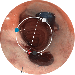
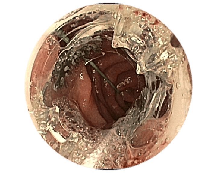
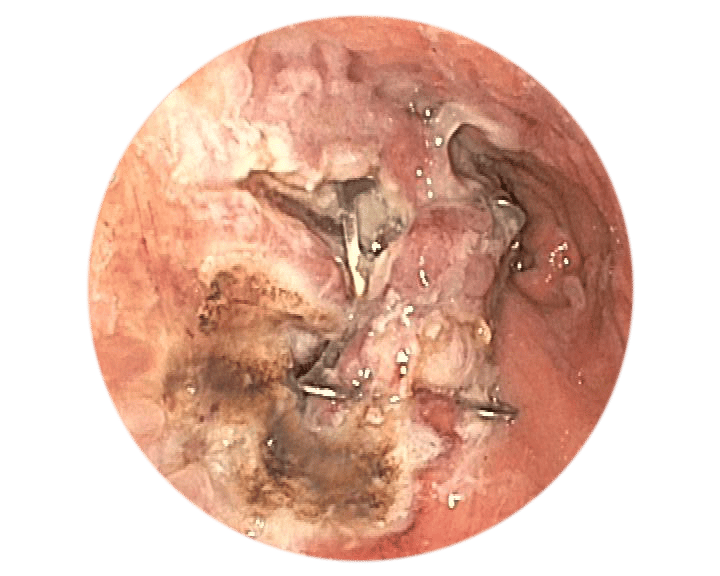
Lumen reduction for bariatric endoscopy

LATEST PRODUCTS

+49 (0) 7071 96528 160
service@ovesco.com

Use of the BARS®

BARS® is an endoscopic treatment of weight regain or dumping syndrome after a gastric bypass.

The BARS®

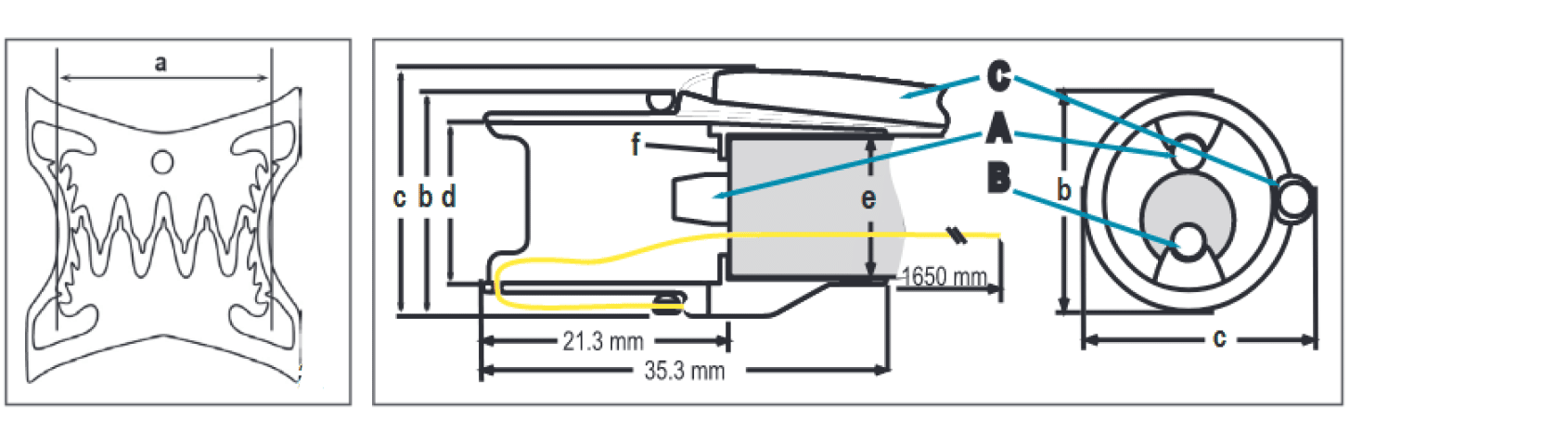
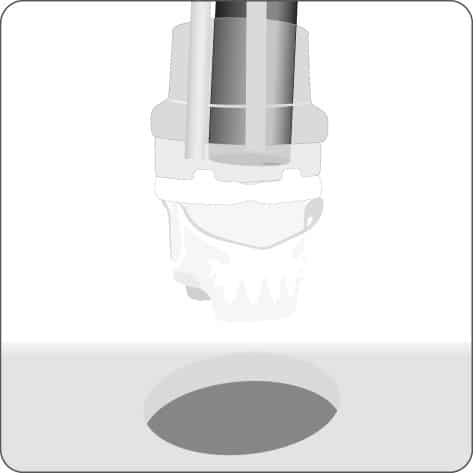
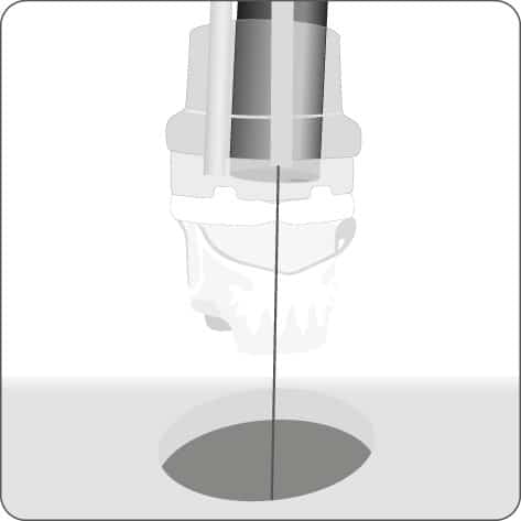
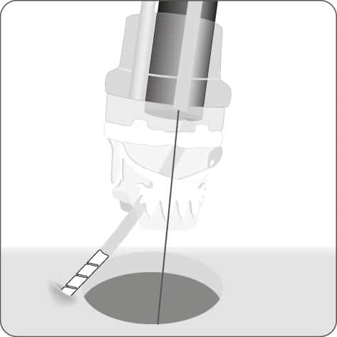
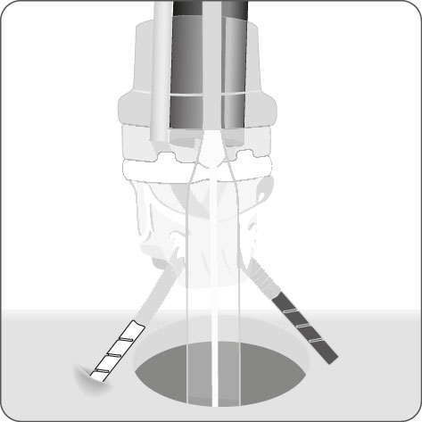
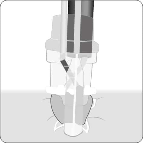
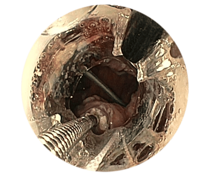
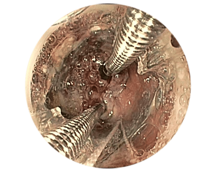
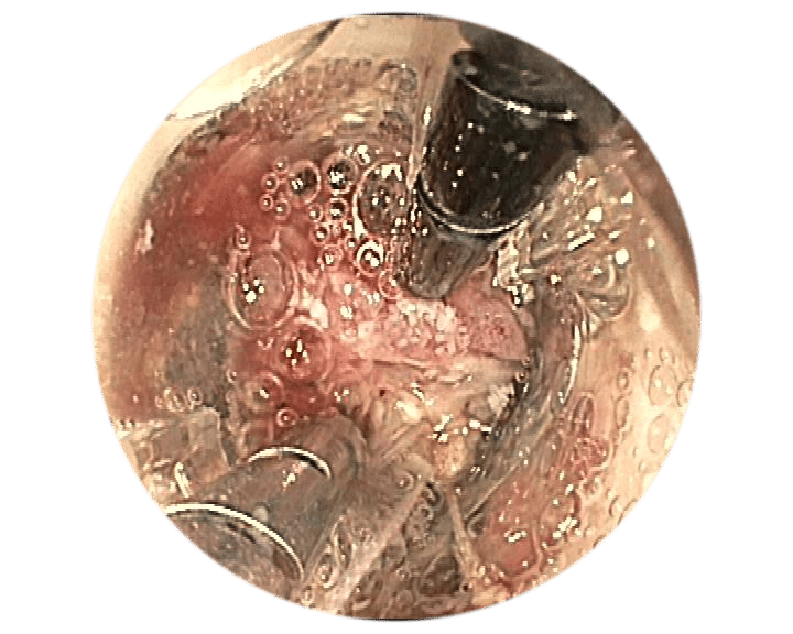
The BARS® consists of an application cap with mounted clip and thread, thread retriever and BARS® hand wheel. The BARS® application cap is mounted on the tip of the endoscope while the application aids are guided along the outside of the endoscope in working channels. By turning the hand wheel, the thread is tensioned and the clip is released.
During clip application, the balloon that is inserted into the anastomosis prevents the lumen from closing completely and defi nes the remaining lumen.

The BARS® Set

The BARS® Set is supplied as a complete treatment unit and comprises the following products:

  • BARS® application cap with mounted clip and thread
  • Two additional working channels that are integrated in the cap
  • BARS® Anchor (1x Silver & 1x Black)
  • BARS® hand wheel
  • Insertion balloon
  • Guide wire
  • Thread retriever
  • Space keeper balloon

Before purchasing and using the BARS®, participation in a training course is mandatory.

Dimensions and specifications

Cap length 35.3 mm
Clip width = compression width [a] 12.4 mm
Min. outer diameter [b] 19.5 mm
Max. outer diameter [c] 21.2 mm
Cap inner diameter [d] 13.1 mm
Compatible endoscope diameter [e] 10.0–12.0 mm
Inner BARS working channel [A] 3.0 mm working channel diameter | Max. 2.8 mm instrument diameter
Endoscope working channel [B] min. 3.7 mm
Outer BARS working channel [C] 2.8 mm working channel diameter | 2.6 mm instrument diameter
Guide wire 3700 mm length | 0.76 mm (0.03‘‘) diameter
Reference number 100.60

Application of the BARS®

EMR2

Mucosal incision2

Preparing the target tissue

Preparing the target tissue using EMR or mucosal
incision offers significant benefits.

  • Enables a more secure hold for the Anchors in the muscular
    (submucosal) layer of tissue.
  • A fresh lesion is created which can promote healing and
    thus support the long-term results.

Positioning

Correct positioning of the BARS® Anchors is crucial to the success of the treatment.

  1. Divide the anastomosis into two halves with an imaginary centre line.
    X = exit point of the endoscope working channel
  2. Position the Anchors just below the centre line.
    • = BARS® Anchor Black, = BARS® Anchor Silver.
  3. The calibration balloon () defines the remaining lumen
    in the opposite semi-circle.

2Source: Prof. A. Schmidt, Universitätsklinikum Freiburg, Germany

Target the application site.

Insert the guide wire into the outer BARS® working channel and place it in the anastomosis.

Insert the anchors into the endoscope working channel and the inner
BARS® working channel.

Crossed placement of the anchors in the prepared target tissue.

Place the calibration balloon in the anastomosis. Gradually pull the tissue into the BARS® application cap using alternating pulling motions.

Make sure that the tissue is positioned symmetrically in the cap. Position the BARS® clip by turning the hand wheel.

Withdraw the instruments and inspect the clip application site.

Example 1*

Target the application site.

Insert the guide wire into the outer BARS® working channel and place it in the anastomosis.

Insert the anchors into the endoscope working channel and the inner
BARS® working channel.

Crossed placement of the anchors in the prepared target tissue.

Place the calibration balloon in the anastomosis. Gradually pull the tissue into the BARS® application cap using alternating pulling motions.

Make sure that the tissue is positioned symmetrically in the cap. Position the BARS® clip by turning the hand wheel.

Withdraw the instruments and inspect the clip application site.

1Source: Dr. med. M. Kandler, Städtisches Klinikum Dresden, Germany

Media

{{pluginSettings.generalSettings.writing.no_results}}
of {{actionSettings.pagesCount}}

TOP