
 Deutsch
  Français
  Español
  Português
 OTSC® application aid for improved mobilisation of the tissue, also in case of scarring of the tissue.


+49 (0) 7071 96528 160
service@ovesco.com
+49 (0) 7071 96528 160
service@ovesco.com
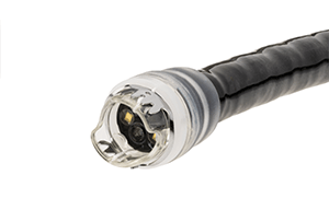
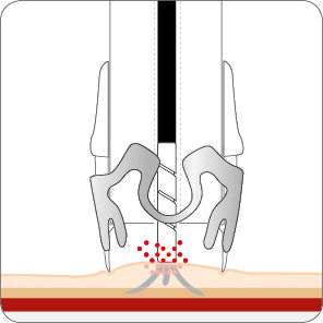
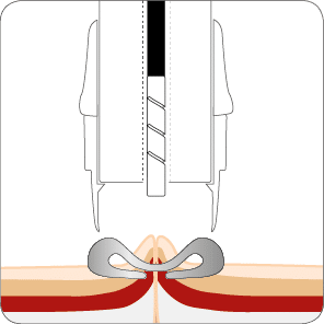
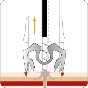
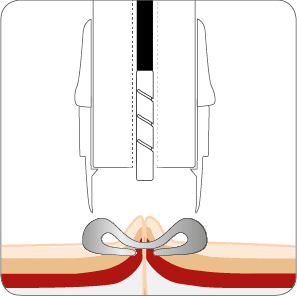
The OTSC® Anchor can be used for better approximation of tissue especially when indurated (e.g. fistulae, chronic ulcer). It also facilitates targeting of lesions, e.g. in the treatment of hemorrhage, the OTSC® Anchor allows precise alignment between the target tissue and the applicator cap.
The OTSC® Anchor is available in two versions and lengths:
| OTSC® Anchor | OTSC® Anchor 220tt | |
|---|---|---|
 |
 |
|
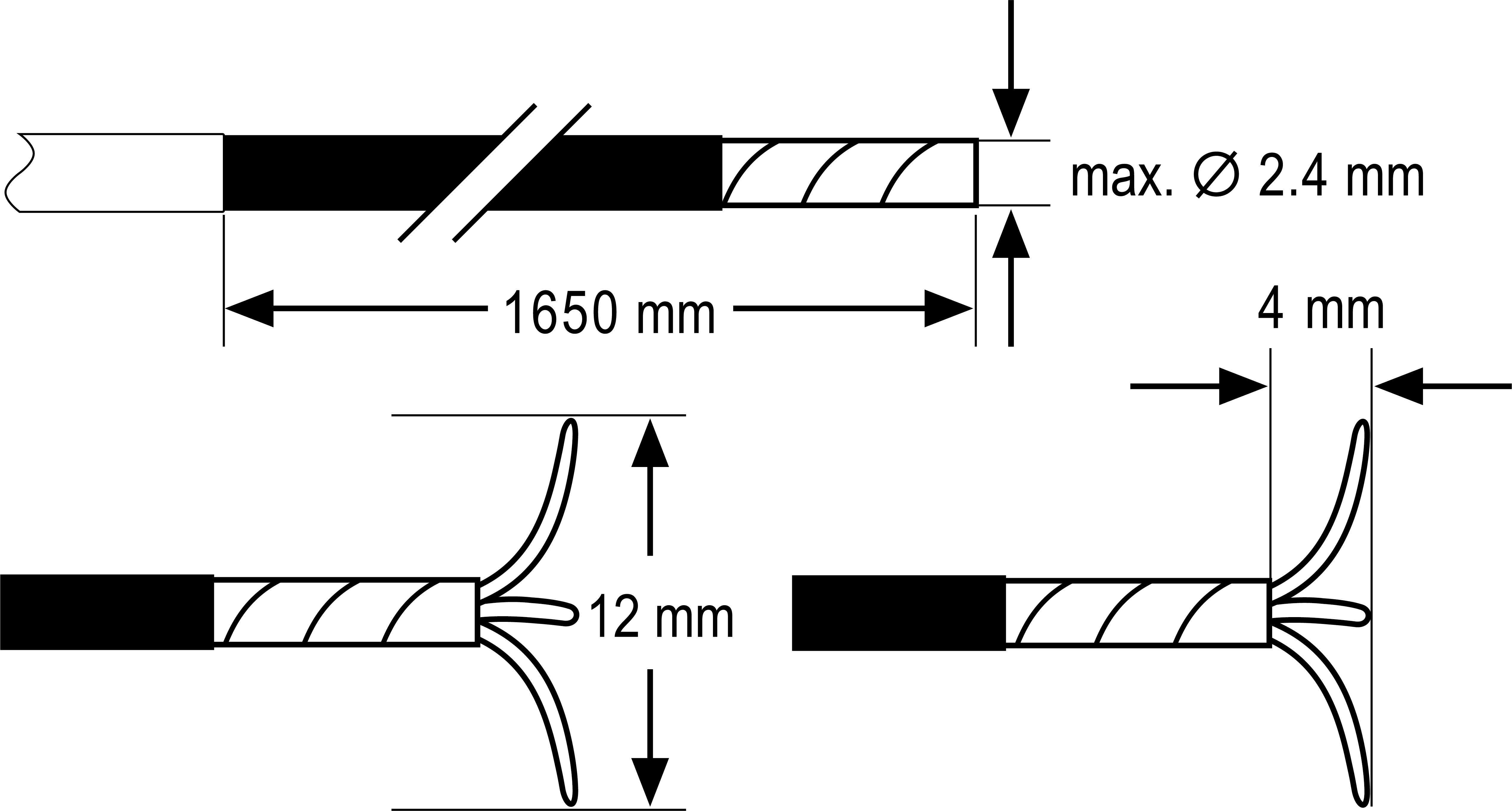
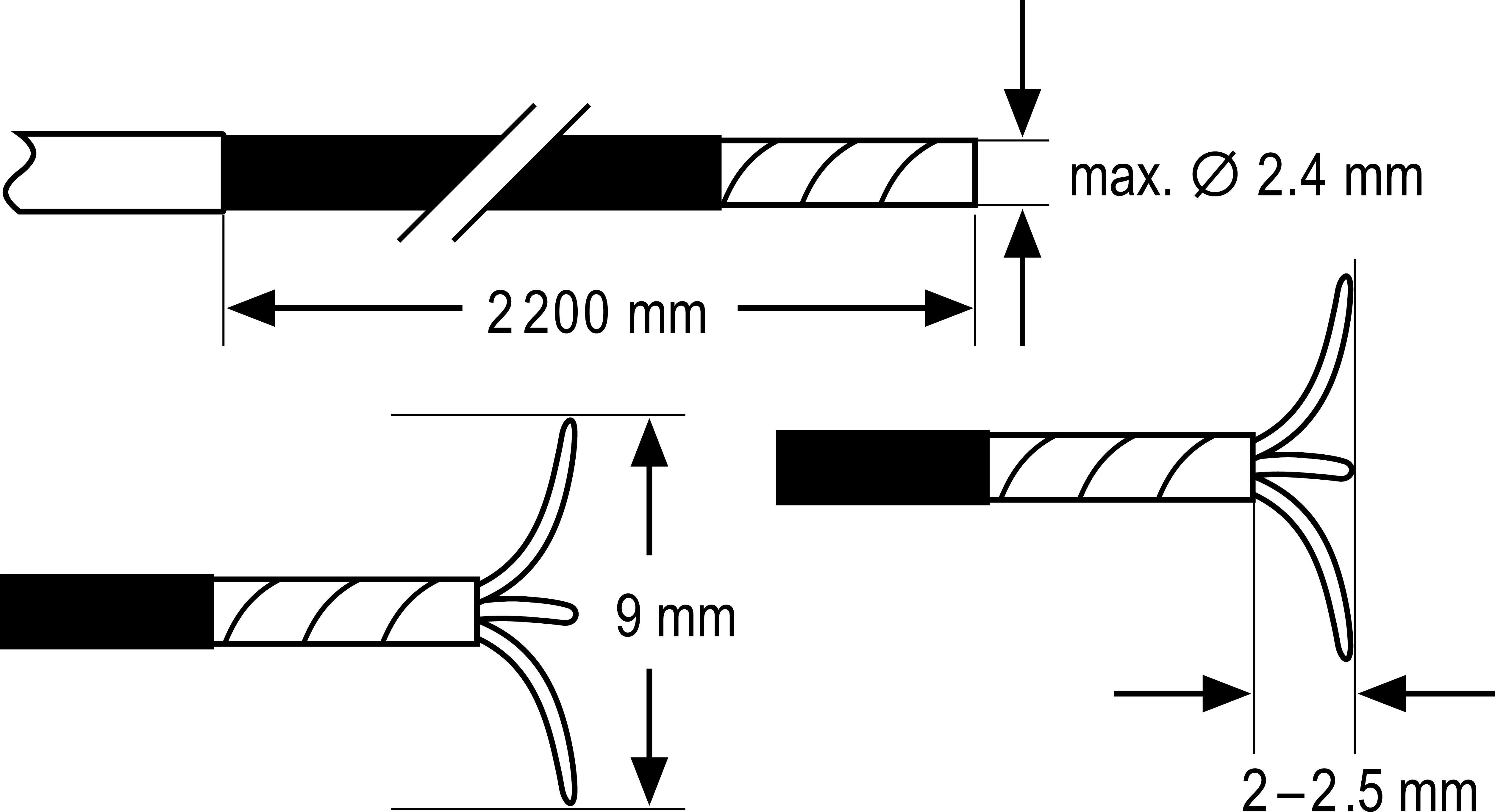
| Needle width | 12 mm | 9 mm |
| Stich depth | 4 mm | 2 – 2.5 mm |
| Length | 165 cm | 220 cm |
| Appropriate endoscope | Working channel diameter of minimum 2.8 mm to be used alone or 3.2 mm with the OTSC® System Set. | Working channel diameter of minimum 2.8 mm to be used alone or 3.2 mm with the OTSC® System Set. |
| Items per package | 1 | 1 |
| Reference number | 200.10 | 200.11 |
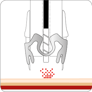
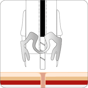
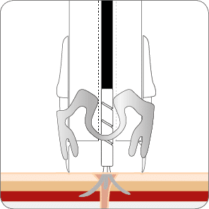
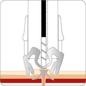
In cases of fibrotic or hard tissue (e.g. callous ulcers) or tangential application, the OTSC® Anchor can be valuable in precisely aligning target tissue with the cap opening and keeping it fixed during clip release. It may not always be possible to manipulate fibrotic tissue fully inside the cap. However, it is sufficient to pull the tissue firmly to the rim of the cap with the OTSC® Anchor, then apply the clip. The clip “jumps” slightly forward upon release and grasps the tissue in front of the cap.


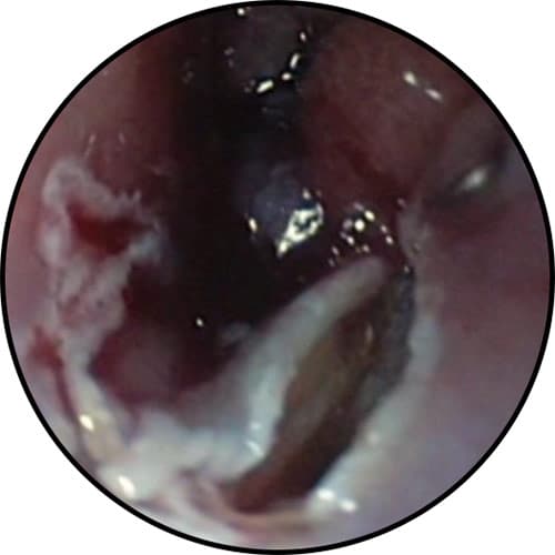
Closure of persistent PEG-fistula using the OTSC® Anchor


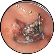
* Source: Dr. Thomas Kratt, Interventional Endoscopy, Klinik für Allgemeine, Viszeral- und Transplantationschirurgie, University Hospital Tuebingen, Germany

Closure of a large esophago-bronchial fistula through mucosal incision prior to OTSC® placement
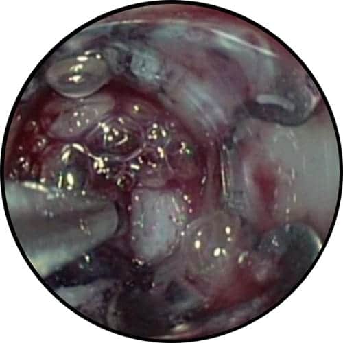

* Source: Meining A. et al. (2015) Erfolgreicher Verschluss einer großen ösophago-bronchialen Fistel durch mukosale Inzision vor OTSC-Klipp-Platzierung. Endoskopie heute. Doi: 10.1055/s-0035-1545049.

Related products