
 Deutsch
  Français
  Español
  Português
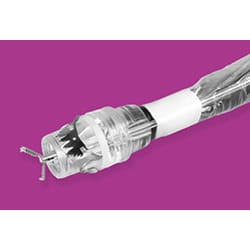
Review on the FTRD® System – Multi-tasker in the clinical routine
Due to its high resection rates, low incidence of adverse events and ease of use, FTRD was quickly established in clinical routine. It is now used in a wide range of indications. The newer gastroduodenal FTRD further expands the scope and is showing promising results in early applications.
The review by Mueller et al., Department of Medicine II, University of Freiburg, Germany, aimed to provide a comprehensive overview on the clinical application and current data of EFTR using the FTRD System.
Indications
The FTRD System is available in several variations for different indications:
- colonic FTRD for use in the colorectum
- diagnostic FTRD for diagnostic resections in the colorectum
- gastroduodenal FTRD for use in the upper GI tract
Colorectal EFTR
For EFTR with the FTRD in the colorectum, the technical success rate ranges from 83.9% to 89.5% and R0 resection from 76.9 % to 82.4%, which is shown by various larger multicenter studies (WALL RESECT study, German colonic FTRD registry and Dutch FTRD registry). The patient population was comprised of various indications for EFTR in the colorectum including a high proportion of T1 carcinoma.
Lesion size is a critical factor in achieving complete resection. In most clinical studies lesion size was ≤ 30 mm. In the presence of scarring or malignant histology, the authors do not recommend selecting lesions > 20 mm for en-bloc EFTR.
Adverse events (AEs) after EFTR in the lower GI tract are reported in 5.3% to 14.1% of cases; the rate of severe AE is in the range of 1.3% to 4.4%. Most frequent complications are bleeding and perforation. While delayed bleeding requiring re-intervention occurs in 0%–6.6% of cases, intraprocedural bleeding at the resection site is very rare. The rate of perforation is about 2.5% (reported in the German FTRD registry and other larger studies). Acute perforation is mainly caused by a wrong sequence of resection steps and can be avoided by proper release of the FTRD clip from the cap (strict visual inspection of the white ring during the procedure). Secondary perforation may be caused by thermal damage and resulting tissue necrosis underneath the clip when an additional snare is used to resect the tissue after failed one-step EFTR. Risk factors such as smoking, body mass index and immunosuppressive therapy may increase the risk of secondary perforation. Further complications can occur because of very rare events like entrapment of adjacent organs into the cap. Most AEs can be managed endoscopically or conservatively; only around 2% of AEs require surgical therapy.
Data on mid- and long-term recurrence is still lacking because the follow-up in most studies does not exceed 3-6 months. The WALL RESECT study shows a recurrence rate of difficult adenomas of 12.3% after 3 months. The German FTRD registry reports a recurrence rate of 13.5% in a median follow-up of 22 weeks (72% occurred after R1/Rx resection and 28% after R0 resection). Several studies showed that the FTRD clip detaches spontaneously in about 70% of cases within 3 months. Treatment options for recurrent lesions after EFTR include re-EFTR, EMR, ESD, or avulsion. In the WALL RESECT study and the German FTRD registry, recurrences could be successfully treated endoscopically in most cases.
EFTR with the FTRD is increasingly used for diagnostic and therapeutic resection of T1 carcinoma, (especially for re-resection in case of R1/Rx resected T1 carcinomas). In addition, EFTR may also be a viable option for treatment-naïve lesions and for patients unsuitable for surgical intervention due to the minimally invasive treatment alternative. High efficacy of EFTR is also shown for subepithelial tumors (SETs) and especially for rectal neuroendocrine tumors (NETs). For lesions at the appendiceal orifice, R0 resection rate differs strongly (from 64% to 93%) with a slightly higher appendicitis rate (15% to 17%). Thus, EFTR may be an alternative to surgery, but patients should be thoroughly informed about the risks of the procedure. Using the novel diagnostic FTRD, EFTR can also be used to obtain full-thickness specimen of the intestinal wall in patients with suspected motility disorders. The device is slightly smaller in size and therefore captures less tissue in the cap. Valli et al. demonstrated that EFTR with the diagnostic FTRD enables precise histological analysis in patients suspected of having neuromuscular gut disorders such as Hirschsprung disease or enteric ganglionitis.
Colorectal EFTR in comparison with EMR and ESD
EFTR for colorectal lesions shows a similar technical success rate to EMR and ESD. Direct comparison with EMR is challenging due to different indications. EMR is typically used for treatment-naive adenomas, while EFTR is applied for non-lifting lesions such as recurrent adenomas. Ongoing research in a multicenter RCT (CURE study) aims to determine if EFTR is superior in reducing recurrences. ESD allows resection of larger lesions with higher en-bloc resection and lower recurrence rates than EMR, but it is technically demanding and has an increased risk of perforation. European centers have gained experience in ESD, and R0 resection rates for FTRD resection are comparable to Western ESD rates.
EFTR in the upper GI tract
The gastroduodenal FTRD is a modified device for use in the upper digestive tract (stomach and duodenum). The system is equipped with a balloon for better passage and is slightly smaller in size than the colorectal version. Studies have shown promising results for resecting subepithelial lesions (SETs) and duodenal adenomas. However, the R0 resection rate was low due to selecting too advanced lesions for en-bloc EFTR. Several studies have demonstrated the feasibility and effectiveness of EFTR for small (≤ 15 mm) SETs in the stomach, but more research is needed to determine its application for duodenal adenomas. Ongoing trials are comparing EFTR with EMR in this context.
Hybrid-EFTR
Hybrid-EFTR is a combination of a conventional EMR followed by EFTR with the FTRD for the remaining lesion and overcomes the size limitation of FTRD resections. First, the lifting parts of the lesion are resected in conventional piecemeal (EMR). Afterwards, the remaining non-lifting central part is removed using EFTR in the same session. Clinical success, technical success and R0 resection rates of Hybrid-EFTR (resection size of 15-70 mm) are similar to the rates of the standalone EFTR (resection size of 7–25 mm). EFTR can also be combined with ESD as shown in the study by Andrisani et al. Here, EFTR with the FTRD was used as a “rescue” procedure after difficult ESD; EFTR increased complete resection rates and reduced complications. Studies on Hybrid-EFTR do not show increased complication rates compared to the standalone technique.
The authors conclude that EFTR using the FTRD is a safe and efficient method for treating lesions that cannot be managed with conventional endoscopic techniques. It is a relatively simple procedure with a steeper learning curve compared to other resection techniques (especially ESD), and it allows for precise histological evaluation, especially in the case of malignant tumors. The development of device design and its combination with other resection techniques have expanded its indications. While EFTR has extensive evidence supporting its efficacy and safety in the colorectum, more research is needed to establish its effectiveness in the upper GI tract.
Mueller J, Kuellmer A, Schiemer M, Thimme R, Schmidt A. Current status of endoscopic full-thickness resection with the full-thickness resection device. Dig Endosc. 2022. doi:10.1111/den.14425.
 |
 |